Transcriptomics
Single-nucleus, single-cell, regional, and spatial transcriptomics: goals, protocols, and metadata standards.
KPMP Transcriptomics technologies
Single-nucleus RNA-seq (snRNA-seq)
Goal: Identify cell types and states associated with normal and injured kidney functions using gene expression profiling. Identify marker genes for cell type/states and any proportion shifts underlying pathology.

Protocols:
Single-cell RNA-seq (scRNA-seq)
Goal: Empirically derive cell subtypes and cell-type-specific gene expression profiles.

Protocol(s)
Regional transcriptomics
Goal: Generate deep transcriptomic signatures from nephron segments defined spatially by antibody staining using laser microdissection.

Protocol(s)
Spatial transcriptomics - Visium
Goal: Whole transcriptome mRNA expression with localization to kidney cells and structures
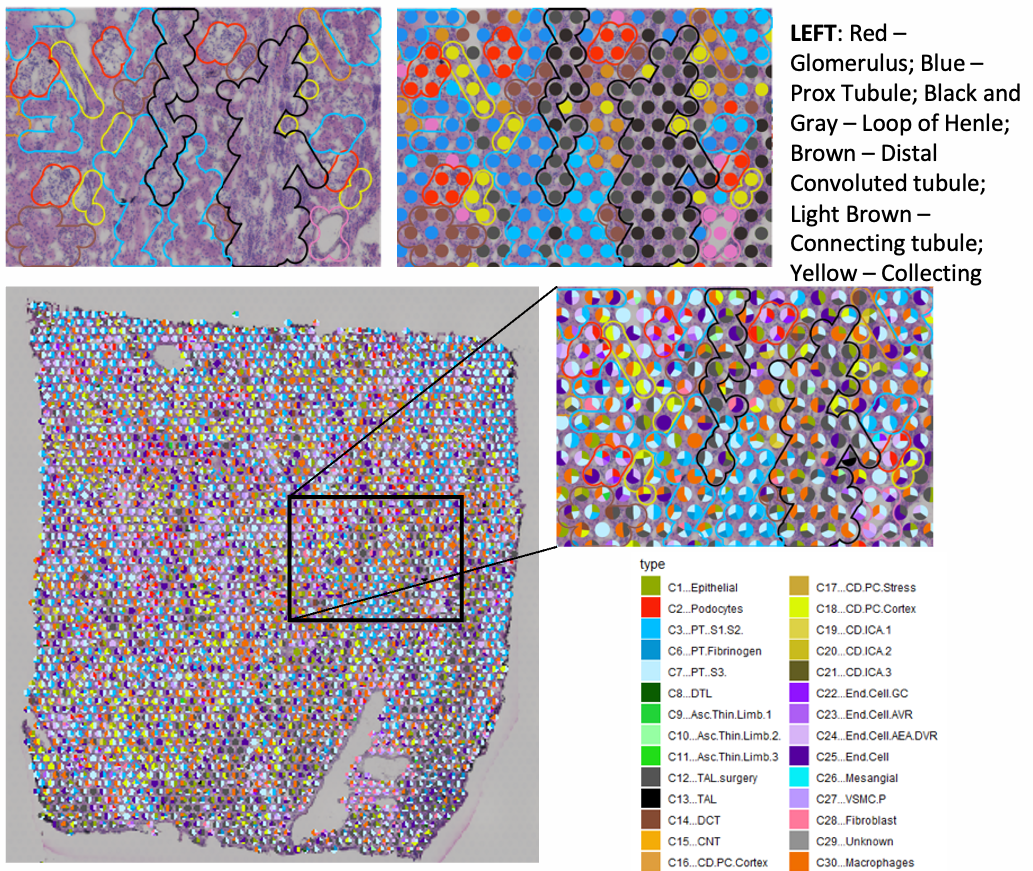
Protocol(s)
Spatial transcriptomics - Xenium
Spatial transcriptomics data generated using the 10x Genomics Xenium platform allows researchers to see where genes are active within a kidney tissue sample. By mapping gene activity directly to individual cells and their locations, Xenium provides a detailed view of how cells are organized and interact within the kidney.
This information helps researchers better understand how kidney diseases develop, identify which cells are involved in injury and repair, and discover new targets for treatments. By combining molecular data with tissue structure, Xenium offers a powerful tool for advancing kidney research and precision medicine.
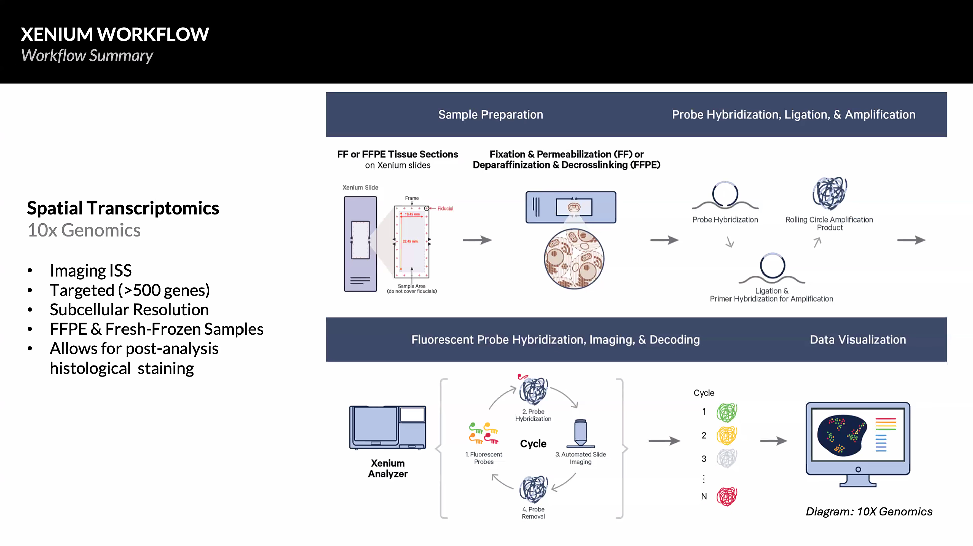
Protocol(s):
Formalin-Fixed Paraffin-Embedded (FFPE)
- Xenium Spatial Transcriptomics with Protein add-on Protocol for Human Tissue (hMulti_480g)
This protocol measures expression of 480 genes, 27 proteins (23 proteins + 4 segmentation markers) and dapi (nuclear staining) defining cellular diversity of healthy and injury states on the same fixed kidney tissue at the same time performed at Washington University.
- Xenium in situ Gene Expression for FFPE – IU KPMP TIS (hKidney_480g)
This is the protocol used by the Indiana University Tissue Interrogation Site for the Kidney Precision Medicine Project consisting of 480 genes, 27 proteins (23 proteins + 4 segmentation markers) and dapi (nuclear staining).
Liquid Nitrogen (LN2) & Carboxymethyl Cellulose (CMC)-Embedded Fresh-Frozen Samples
- 10X Xenium In Situ Gene Expression for Fresh-Frozen Kidney (VU_hKidney_300g) This is a protocol used by the Vanderbilt University Tissue Interrogation Site for preparing fresh-frozen kidney tissue for in situ spatial transcriptomics using a 300 gene custom probe panel with the 10X Xenium Analyzer platform. VU’s panel targets major/minor/rare kidney cell types, immune processes, and biochemical pathways related to cell signaling and lipid biosynthesis.
10X Multiome (snRNA-seq + snATAC-seq)
Goal: 10X Genomics multiome protocol generates a molecular atlas of the human kidney with comprehensive cell types and minimal processing artifacts. 10X Multiome snATAC-seq + Gene Expression assay allows measurements of chromatin accessibility and transcription from the same cell. This allows direct insights into putative transcription factors or SNPs associated with open chromatin and impact on gene expression.
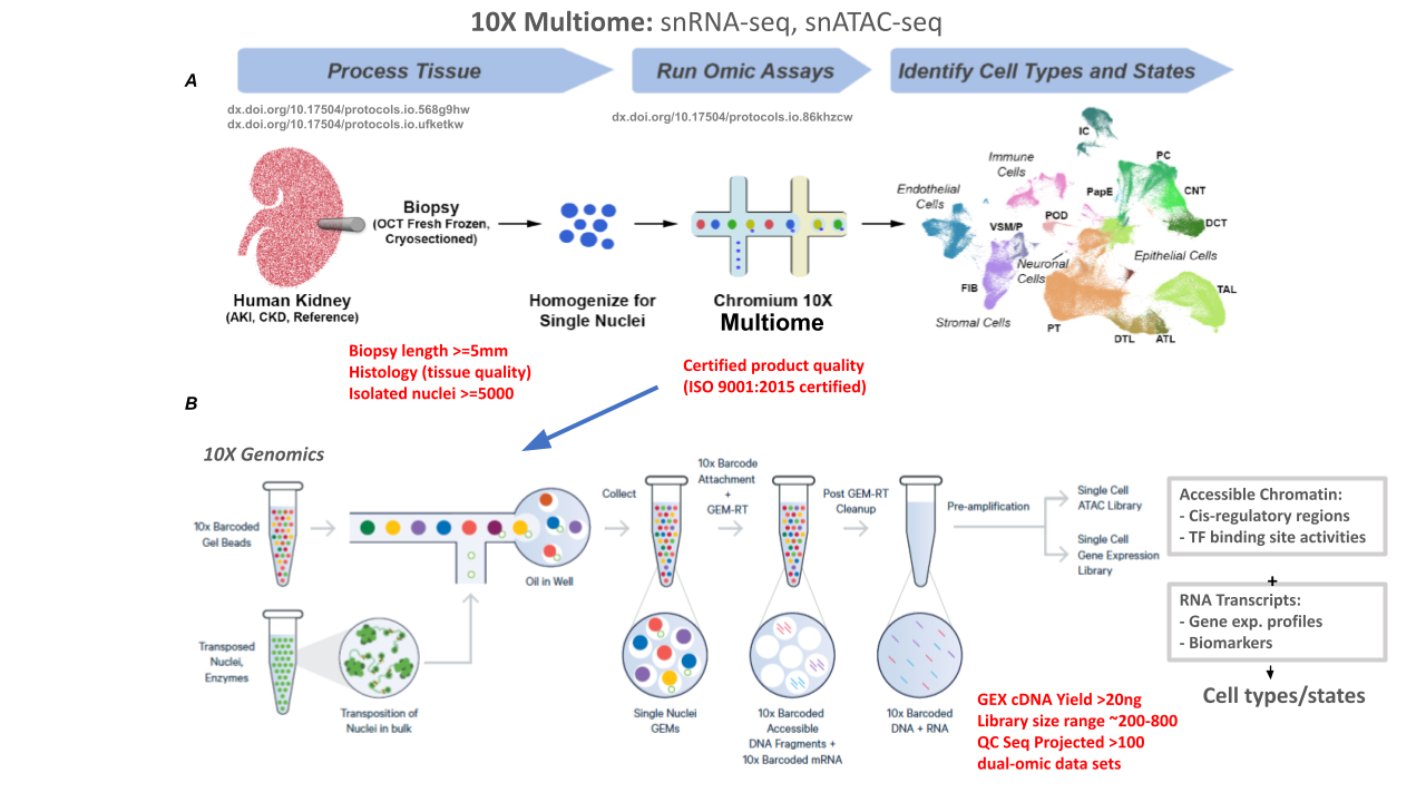
Protocol(s):
Metadata standards